Hernia anatomy of the muscles, arteries, veins, and nerves of the abdominal wall, especially within the groin and inguinal region, are some of the most difficult to understand – even for doctors! However, learning which muscles are weakened in a hernia and understanding hernia anatomy can help patients understand where hernias are located, how they form, and how we repair them.
We hope that the following diagrams, pictures, and text help you understand more about the anatomy of an abdominal wall hernia.
Hernia Anatomy – The layers of the Abdominal Wall
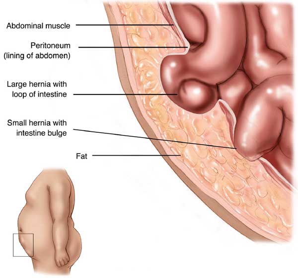
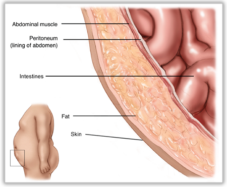 The first concept to understand is the basic layers of the abdominal wall. These layers are a bit different between the umbilical region and the groin, but overall the basic layers are the same. From the outside to the inside is the skin, then a layer of fat. Underneath the fat is the layer of muscles which provide the strength to the abdominal wall. Under the muscles is a thin layer called peritoneum which serves as a barrier between the muscles and the internal organs which live underneath the peritoneum.
Inguinal Hernia Anatomy
In inguinal or groin hernias a hole forms in the internal oblique and transversus muscles. If this hole forms lateral (or away from the middle) to the inferior epigastric blood vessels, an indirect inguinal hernia forms. If the hole forms medial (or towards the middle) to the inferior epigastric blood vessels, a direct inguinal hernia is formed. Regardless, in open surgery, the external oblique muscle layer is opened over the hernia (weakened internal and transversus muscle). The hole (or holes) in the internal oblique and the transversus muscle are found. A dual sided mesh is used to reinforce the hernia defect and the muscle around the hole. 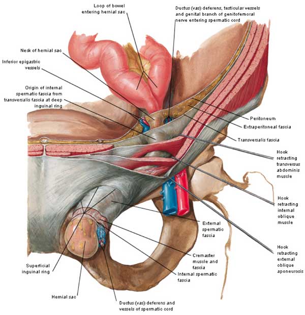
The image below shows the anatomy of the inguinal region looking from the inside of the body outwards. This would be the view from inside the right groin. The right ‘indirect’ hernia is lateral or away from the middle of the patient. The left ‘direct’ hernia is medial or towards the middle compared to the inferior epigastric vessels. 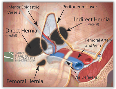
Umbilical Hernia Anatomy
The CT image below shows a cross section of a patient. You can see a clear hole in the muscle at the level of the belly button, or umbilicus. This hole is considered an umbilical hernia. 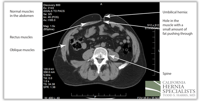 The image below shows a normal umbilicus with no evidence of a hole, or a hernia. Although CT scans can be helpful in determining whether there is a hernia, most often examining the patient is all that is needed to determine whether there is a hernia present.
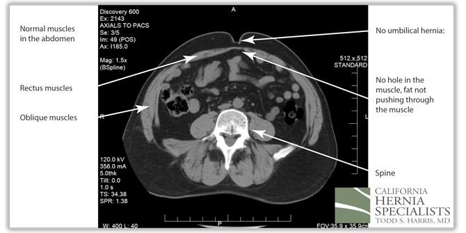
Tension Free Repair
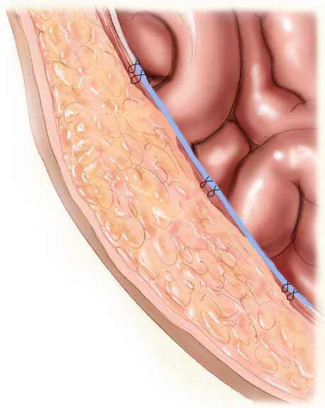
The term ‘tension free’ hernia repair is commonly used to describe hernia surgery. Hernias are caused by a weakening of the abdominal muscles. Some surgeons choose to sew the muscles back together, thus causing ‘tension’ on the muscles around the hernia. However, the muscles around a hernia are already weak, and over time those muscles tend to pull apart and the hernia can recur, or come back. Therefore, most hernia specialists today utilize a mesh to help strengthen the muscles. When using a mesh, the muscles themselves are not sewn together (see below). Instead, a mesh is placed over or under the hole in the muscle to prevent anything from pushing through the abdominal wall.
Some patients have heard or read negative information about mesh surgery. However, the unbiased government agency The National Institutes of Health performed a study of whether mesh should or should not be used for hernia surgery. Read the article by clicking on the logo to the right:
While there are some risks with any surgery, by utilizing the newest mesh available, these risks are minimized. Patients can read more on our ‘Truths About Hernia Mesh‘ page.
Our Approach
Dr. Harris specializes in surgical repair for all hernia types. For patients who require a laparoscopic surgery, Dr. Harris commonly performs these procedures. When performing a tension free mesh hernia repair, Dr. Harris uses the latest and most advanced lightweight mesh available which significantly reduces pain and discomfort after the surgery. Many surgeons still use mesh which was invented 10 years ago which can increase the chances of post operative mesh pain. Today’s advanced mesh reduces those risks to almost zero.
Although open surgical hernia repair is still an important option for many patients, every patient should consider being seen by a surgeon who can perform advanced laparoscopic hernia surgery and who uses the newest lightweight mesh. Only a small handful of the hundreds of general surgeons in Orange County, Los Angeles, Riverside and San Diego have the experience to perform hernia surgeries using laparoscopic techniques and use cutting edge lightweight mesh.
Dr. Harris sees patients from all over Southern California, the greater US, and Canada. Our Newport Beach office is conveniently located in Central Orange county adjacent to John Wayne Airport between the 405, 73, and 55 highways. Our office staff are experienced in verifying insurance coverage for each patient, as well as offering excellent pricing for patients without insurance (see ‘Costs‘ page). We are happy to discuss all forms of payments with patients as needed.


