Specialist Overview: The Mechanics of the Abdominal Wall
Understanding hernia anatomy requires a look at the “inguinal canal”—a natural tunnel through the layers of the abdominal wall. In males, this canal carries the spermatic cord, while in females, it supports the round ligament. A hernia occurs when the integrity of these muscular layers fails, allowing internal tissue to protrude through a weak point.- Direct vs. Indirect: An indirect hernia follows the “official” tunnel of the inguinal canal (often a congenital weakness), whereas a direct hernia punches through a weak spot in the posterior wall known as Hesselbach’s Triangle.
- The Myopectineal Orifice: This is the collective “weak zone” of the groin where inguinal, femoral, and obturator hernias occur.
- Fascial Integrity: The strength of your repair depends on the health of the transversalis fascia and the surrounding musculature, which we evaluate to determine if a no-mesh or mesh-reinforced repair is most appropriate.
Anatomic Precision in Surgery
At California Hernia Specialists, our high-volume experience allows for a sophisticated understanding of these anatomic variations. Led by Dr. Todd Harris, an 11-year OCMA Physician of Excellence (2015–2025), we focus on “Anatomic Restoration”—returning your structures to their original, healthy orientation. Our surgical protocols are backed by national research (NCT05929937) and published data on patient outcomes (PMID: 39724506).Hernia Anatomy – The layers of the Abdominal Wall
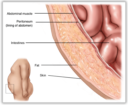 The first concept to understand is the basic layers of the abdominal wall. These layers are a bit different between the umbilical region and the groin, but overall the basic layers are the same. From the outside to the inside is the skin, then a layer of fat. Underneath the fat is the layer of muscles which provide the strength to the abdominal wall. Under the muscles is a thin layer called peritoneum which serves as a barrier between the muscles and the internal organs which live underneath the peritoneum.
Inguinal Hernia Anatomy
In inguinal or groin hernias a hole forms in the internal oblique and transversus muscles. If this hole forms lateral (or away from the middle) to the inferior epigastric blood vessels, an indirect inguinal hernia forms. If the hole forms medial (or towards the middle) to the inferior epigastric blood vessels, a direct inguinal hernia is formed. Regardless, in open surgery, the external oblique muscle layer is opened over the hernia (weakened internal and transversus muscle). The hole (or holes) in the internal oblique and the transversus muscle are found. A dual sided mesh is used to reinforce the hernia defect and the muscle around the hole.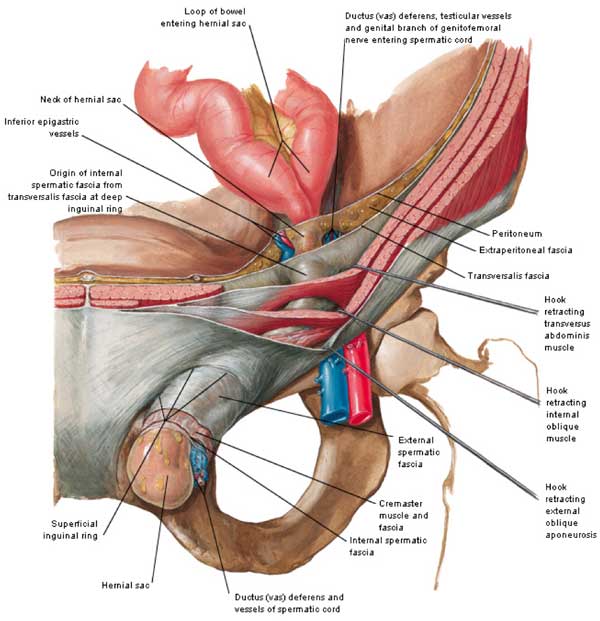 The image below shows the anatomy of the inguinal region looking from the inside of the body outwards. This would be the view from inside the right groin. The right ‘indirect’ hernia is lateral or away from the middle of the patient. The left ‘direct’ hernia is medial or towards the middle compared to the inferior epigastric vessels.
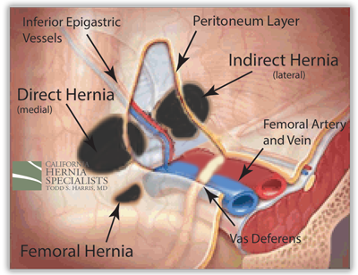
Umbilical Hernia Anatomy
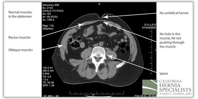
The CT image below shows a cross section of a patient. You can see a clear hole in the muscle at the level of the belly button, or umbilicus. This hole is considered an umbilical hernia.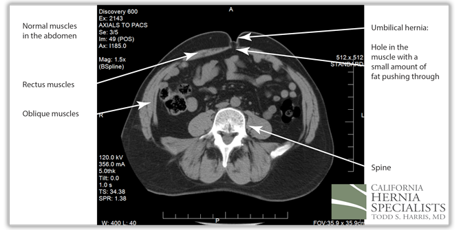 The image below shows a normal umbilicus with no evidence of a hole, or a hernia. Although CT scans can be helpful in determining whether there is a hernia, most often examining the patient is all that is needed to determine whether there is a hernia present.